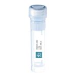
MOG (91-108) peptide- Myelin Oligodentrocyte Glycoprotein peptide 91-108,Mouse
MOG 91-108: SDEGGYTCFFRDHSYQEE
MOG (Myelin Oligodendrocyte Glycoprotein – UniProtKB – Q61885) is a protein with a molecular weight of around 26-28 kDa,located on the external surface of the peripheral lamellae of myelin sheaths of the CNS (Central Nervous System). MOG is an important antigenic target in autoimmune diseases that mediates demyelination in the CNS.
MOG91-108 induces EAE
This peptide is a fragment of amino acids 91-108 from Myelin oligodendrocyte glycoprotein (MOG). MOG (91-108) peptide contributes to multiple signaling pathways in lymphocytes as well as in organ-resident cells like in the CNS. MOG 91-108 is a one of the major determinant to trigger disease in rats expressing RT1av1 or RT1n haplotypes. Moreover,in MOG 91-108-induced EAE (Experimental Autoimmune Encephalomyelitis) the interaction of CXCR4 and CXCL12 is of paramount importance for disease development. Therefore MOG91-108 upregulate the activity of the chemokine receptor CXCR4 and its ligand CXCL12.

 Click here to discover SB-PEPTIDE’s Human MOG peptides pool for T-cells activation
Technical specification
 |
Sequence : SDEGGYTCFFRDHSYQEE – Purity : > 95% |
 |
MW : 2170.19 g / mol (C93H124N24O35S) |
 |
For research use |
 |
Counter-Ion : TFA Salts (see option TFA removal) |
 |
Delivery format : Freeze dried in polypropylene 2mL microtubes |
 |
Other names : Myelin Oligodendrocyte Glycoprotein (91-108) |
 |
Peptide Solubility Guideline |
 |
Bulk peptide quantities available |
Price
| Product catalog | Size | Price € HT | Price $ USD |
| SB141-1MG | 1 mg | 110 | 138 |
| SB141-5MG | 5 mg | 385 | 481 |
References
J. Immunol 2001 doi: 10.4049/jimmunol.166.12.7588.
MHC class II-regulated central nervous system autoaggression and T cell responses in peripheral lymphoid tissues are dissociated in myelin oligodendrocyte glycoprotein-induced experimental autoimmune encephalomyelitis
We dissected the requirements for disease induction of myelin oligodendrocyte glycoprotein (MOG)-induced experimental autoimmune encephalomyelitis in MHC (RT1 in rat) congenic rats with overlapping MOG peptides. Immunodominance with regard to peptide-specific T cell responses was purely MHC class II dependent,varied between different MHC haplotypes,and was linked to encephalitogenicity only in RT1.B(a)/D(a) rats. Peptides derived from the MOG sequence 91-114 were able to induce overt clinical signs of disease accompanied by demyelinated CNS lesions in the RT1.B(a)/D(a) and RT1(n) haplotypes. Notably,there was no detectable T cell response against this encephalitogenic MOG sequence in the RT1(n) haplotype in peripheral lymphoid tissue. However,CNS-infiltrating lymphoid cells displayed high IFN-gamma,TNF-alpha,and IL-4 mRNA expression suggesting a localization of peptide-specific reactivated T cells in this compartment. Despite the presence of MOG-specific T and B cell responses,no disease could be induced in resistant RT1(l) and RT1(u) haplotypes. Comparison of the number of different MOG peptides binding to MHC class II molecules from the different RT1 haplotypes suggested that susceptibility to MOG-experimental autoimmune encephalomyelitis correlated with promiscuous peptide binding to RT1.B and RT1.D molecules. This may suggest possibilities for a broader repertoire of peptide-specific T cells to participate in disease induction. We demonstrate a powerful MHC class II regulation of autoaggression in which MHC class II peptide binding and peripheral T cell immunodominance fail to predict autoantigenic peptides relevant for an autoaggressive response. Instead,target organ responses may be decisive and should be further explored.
Dis Model Mech. 2016 Oct 1;9(10):1211-1220. doi: 10.1242/dmm.025536. Epub 2016 Aug 12.
Identification of gene expression patterns crucially involved in experimental autoimmune encephalomyelitis and multiple sclerosis
After encounter with a central nervous system (CNS)-derived autoantigen,lymphocytes leave the lymph nodes and enter the CNS. This event leads only rarely to subsequent tissue damage. Genes relevant to CNS pathology after cell infiltration are largely undefined. Myelin-oligodendrocyte-glycoprotein (MOG)-induced experimental autoimmune encephalomyelitis (EAE) is an animal model of multiple sclerosis (MS),a chronic autoimmune disease of the CNS that results in disability. To assess genes that are involved in encephalitogenicity and subsequent tissue damage mediated by CNS-infiltrating cells,we performed a DNA microarray analysis from cells derived from lymph nodes and eluted from CNS in LEW.1AV1 (RT1av1) rats immunized with MOG 91-108. The data was compared to immunizations with adjuvant alone or naive rats and to immunizations with the immunogenic but not encephalitogenic MOG 73-90 peptide. Here,we show involvement of Cd38 , Cxcr4 and Akt and confirm these findings by the use of Cd38-knockout (B6.129P2-Cd38tm1Lnd/J) mice,S1P-receptor modulation during EAE and quantitative expression analysis in individuals with MS. The hereby-defined underlying pathways indicate cellular activation and migration pathways mediated by G-protein-coupled receptors as crucial events in CNS tissue damage. These pathways can be further explored for novel therapeutic interventions.
