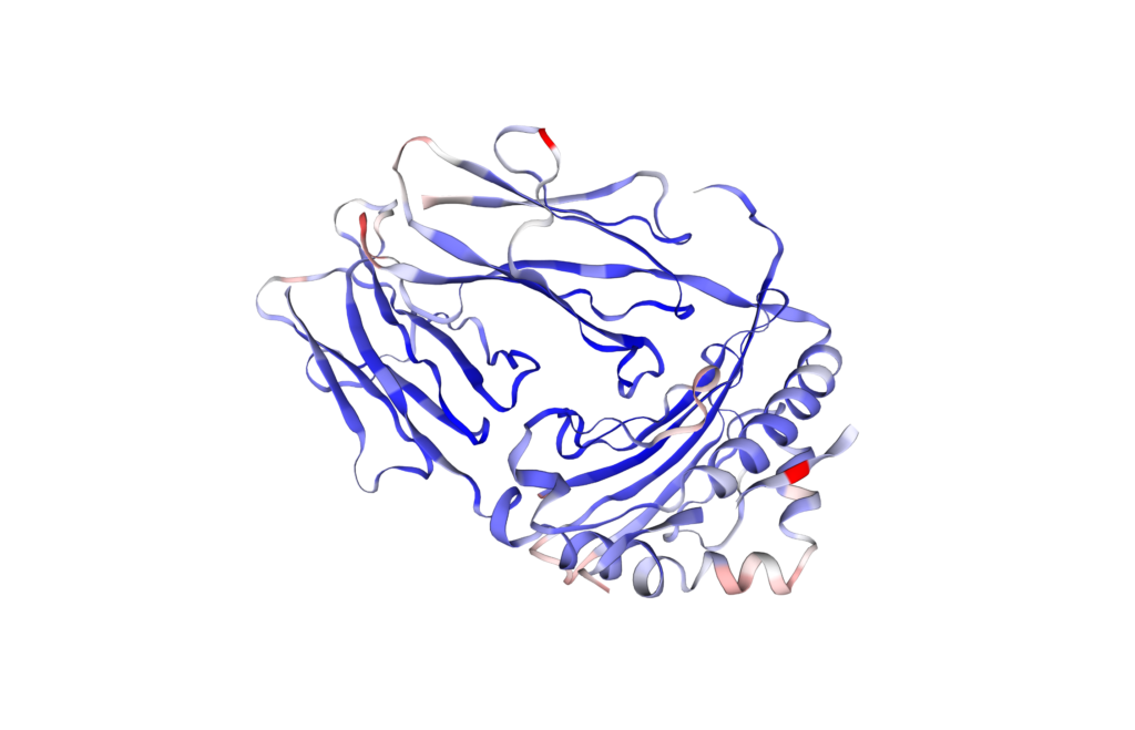
Melan-A (26-35) peptide (EAAGIGILTV) – Melanoma-Associated Antigen
Native Melan-A (26-35) decapeptide derives from the melanocyte lineage-specific protein Melan-A/MART-1 (UniProtKB – Q16655),which is expressed in almost 75-100% of primary and metastatic melanomas.
Melan-A/MART-1 protein
Melan-A,also known as melanoma antigen,recognized by T-cells 1 (Th1) or MART-1 cells,is a 118 amino acids (AA) type III transmembrane protein,consisting in a 26 AA extracellular region (AA 1-26) and a 71 AA cytoplasmic domain.

The latter is only expressed on the surface of melanocytes,melanoma,and retinal pigment epithelium cells,but not in other normal tissues. This unique feature is used in anatomic pathology for melanocytic differentiation that is useful to diagnose melanomas.
The latter is found in the Golgi apparatus but the inversion of its membrane topology leads to its retention in the endoplasmic reticulum (ER). Human Melan-A is involved in melanosome biogenesis by maintaining the stability of GPR143 and its vital role in the functioning of glycoprotein-100 (gp100),also known as the melanocyte protein (PMEL),which is a critical player in the formation of stage II melanosomes.
[CAS: 156251-01-3] – Melan-A (26-35) antigenic peptide
The region 26-35 of Melan-A protein acts as an antigenic peptide that is recognized by CD8+ tumor-reactive cytolytic T lymphocytes (CTLs) for designing antigen-specific cancer vaccines. It has been shown that CD8+ Melan-A-specific CTLs isolated from melanoma patients efficiently lyse the Melan-A-expressing HLA-A*0201 melanoma cell line. However,CTLs preferentially recognize the Melan-A (26-35) peptide as compared with the Melan-A (27-35) peptide.
It has been reported that the concentration of Melan-A (26-35) required to obtain 50% of maximal antigenic activity is EC50 = 0.25 nM and that 20 nM was enough to achieve 50% inhibition of tumor lysis (IC50). Moreover,the high stability of Melan-A (26-35) peptide in human serum (t1/2 = 45 min rather than 5 min for the Melan-A (27-35) nonapeptide) placed Melan-A (26-35) peptide as an antigen of choice for promising cancer vaccines development.
SB-PEPTIDE also offers the A27L version of Melan-A (26-35) peptide,as well as its scrambled version (see sections « Melan-A (26-35) A27L peptide » & « Melan-A (26-35) scrambled »).
Technical specification
 |
Sequence : EAAGIGILTV |
 |
MW : 943.10 g/mol (C42H74N10O14) |
 |
Purity : > 95% |
 |
Counter-Ion : TFA Salts (see option TFA removal) |
 |
Delivery format : Freeze dried in propylene 2mL microtubes |
 |
Peptide Solubility Guideline |
 |
Bulk peptide quantities available |
Price
| Product catalog | Size | Price € HT | Price $ USD |
| SB026-1MG | 1 mg | 61 | 76 |
| SB026-5MG | 5 mg | 209 | 261 |
| SB026-2*5MG | 2×5 mg | 297 | 371 |
| SB026-5*10MG | 5×10 mg | 561 | 701 |
References
Frontiers. 2022 Apr 12;9:873777. doi: https://doi.org/10.3389/fmolb.2022.873777
The Many Faces of G Protein-Coupled Receptor 143,an Atypical Intracellular Receptor
GPCRs transform extracellular stimuli into a physiological response by activating an intracellular signaling cascade initiated via binding to G proteins. Orphan G protein-coupled receptors (GPCRs) hold the potential to pave the way for development of new , innovative therapeutic strategies. In this review we will introduce G protein- coupled receptor 143 (GPR143),an enigmatic receptor in terms of classification within the GPCR superfamily and localization. GPR143 has not been assigned to any of the GPCR families due to the lack of common structural motifs. Hence we will describe the most important motifs of classes A and B and compare them to the protein sequence of GPR143. While a precise function for the receptor has yet to be determined,the protein is expressed abundantly in pigment producing cells. Many GPR143 mutations cause X-linked Ocular Albinism Type 1 (OA1 , Nettleship-Falls OA) , which results in hypopigmentation of the eyes and loss of visual acuity due to disrupted visual system development and function. In pigment cells of the skin,loss of functional GPR143 results in abnormally large melanosomes (organelles in which pigment is produced). Studies have shown that the receptor is localized internally,including at the melanosomal membrane,where it may function to regulate melanosome size and/or facilitate protein trafficking to the melanosome through the endolysosomal system. Numerous additional roles have been proposed for GPR143 in determining cancer predisposition,regulation of blood pressure,development of macular degeneration and signaling in the brain,which we will briefly describe as well as potential ligands that have been identified. Furthermore,GPR143 is a promiscuous receptor that has been shown to interact with multiple other melanosomal proteins and GPCRs,which strongly suggests that this orphan receptor is likely involved in many different physiological actions.
J. Immunol. 2001 Nov 15;167(10):5852-61. doi: 10.4049/jimmunol.167.10.5852
A new generation of Melan-A/MART-1 peptides that fulfill both increased immunogenicity and high resistance to biodegradation: implication for molecular anti-melanoma immunotherapy
Intense efforts of research are made for developing antitumor vaccines that stimulate T cell-mediated immunity. Tumor cells specifically express at their surfaces antigenic peptides presented by MHC class I and recognized by CTL. Tumor antigenic peptides hold promise for the development of novel cancer immunotherapies. However,peptide-based vaccines face two major limitations: the weak immunogenicity of tumor Ags and their low metabolic stability in biological fluids. These two hurdles,for which separate solutions exist,must,however,be solved simultaneously for developing improved vaccines. Unfortunately,attempts made to combine increased immunogenicity and stability of tumor Ags have failed until now. Here we report the successful design of synthetic derivatives of the human tumor Ag Melan-A/MART-1 that combine for the first time both higher immunogenicity and high peptidase resistance. A series of 36 nonnatural peptide derivatives was rationally designed on the basis of knowledge of the mechanism of degradation of Melan-A peptides in human serum and synthesized. Eight of them were efficiently protected against proteolysis and retained the antigenic properties of the parental peptide. Three of the eight analogs were twice as potent as the parental peptide in stimulating in vitro Melan-specific CTL responses in PBMC from normal donors. We isolated these CTL by tetramer-guided cell sorting and expanded them in vitro. The resulting CTL efficiently lysed tumor cells expressing Melan-A Ag. These Melan-A/MART-1 Ag derivatives should be considered as a new generation of potential immunogens in the development of molecular anti-melanoma vaccines.
JBC. 2001 Sept 10;276(46):43189-43196. doi: 10.1074/jbc.M103221200
Subcellular Localization of the Melanoma-associated Protein Melan-AMART-1 Influences the Processing of Its HLA-A2-restricted Epitope
The peptide derived from the melanoma-associated protein Melan-A (Melan-A26 –35/HLA-A2) is an attractive candidate for tumor immunotherapy but little is known about the intracellular processing of this antigen. Here we show that Melan-A is a single-pass membrane protein with an NH2 terminus exposed to the lumen of the exocytic compartment. In transfected melanoma cells,Melan-A accumulates in the Golgi region. Inversion of the membrane topology leads to the retention of Melan-A in the endoplasmic reticulum. Most strikingly,melanoma cells expressing this form of Melan-A are more effectively recognized by specific CTL than those expressing either Melan-A in its native membrane orientation or Melan-A artificially localized in the cytosol. Our data are compatible with the notion that proteins retained in the endoplasmic reticulum are more efficiently degraded and produce more antigenic peptides.
J. Exp. Med. 1999 Sep 20;190(6):775–782. doi: 10.1084/jem.190.6.775
In vivo expression of natural killer cell inhibitory receptors by human melanoma-specific cytolytic T lymphocytes
Natural killer (NK) receptor signaling can lead to reduced cytotoxicity by NK cells and cytolytic T lymphocytes (CTLs) in vitro. Whether T cells are inhibited in vivo remains unknown,since peptide antigen-specific CD8(+) T cells have so far not been found to express NK receptors in vivo. Here we demonstrate that melanoma patients may bear tumor-specific CTLs expressing NK receptors. The lysis of melanoma cells by patient-derived CTLs was inhibited by the NK receptor CD94/NKG2A. Thus,tumor-specific CTL activity may be decreased through NK receptor triggering in vivo.
Curr Opin Immunol.2000 Oct 01;12(5):576-82. doi: 10.1016/s0952-7915(00)00145-x
Design and evaluation of antigen-specific vaccination strategies against cancer
After studies in preclinical mouse models,the efficacy and safety of tumor-specific vaccination strategies is currently being evaluated in cancer patients. The first wave of clinical trials has shown that in general such vaccination strategies are safe. However examples of clinical responses,especially in conjunction with vaccine-induced immune responses,are still scarce. The fact that most trials have so far been performed with end-stage cancer patients can largely account for this deficit. Greater efficacy of anticancer vaccines is expected in patients with less-progressed disease. In addition,the detection of both natural and vaccine-induced T cell immunity needs further improvement.
